
 English

- Welcome
- Ligament injuries
Ligament injuries
##Anatomical_zone##
Introduction
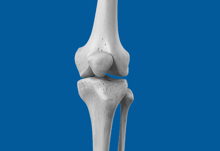
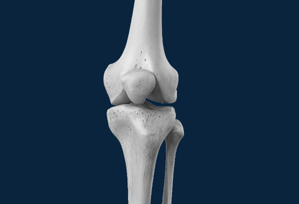
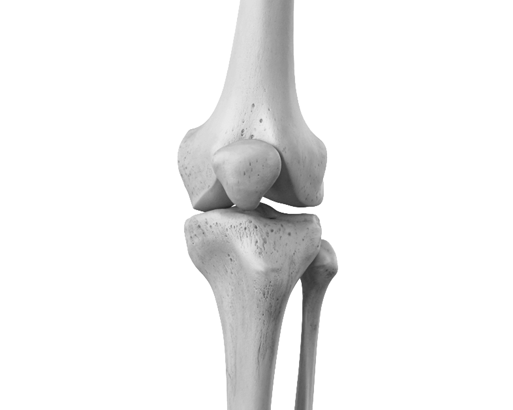
The knee comprises two distinct joints: a weight-bearing condylar joint formed by the femur and tibia, divided into two compartments, medial and lateral, and a trochlear joint formed by the patella and femur. The patella is a sesamoid bone, meaning a bone embedded within a tendon (in this case, the quadriceps tendon). This joint guides the knee's extensor mechanism and amplifies the force of the quadriceps muscle located on the anterior aspect of the thigh.

Do you need the advice of a specialist?
Consult a doctor at the Ouchy Orthopedic Centre.

ACL rupture
Introduction – Epidemiology
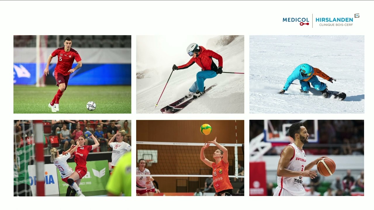
An anterior cruciate ligament (ACL) tear is a common injury in sports. There are currently over 3,000 cases per year in Switzerland (over 200,000 in the USA), and this number continues to rise annually. The incidence is higher in women. This injury frequently occurs during a sudden change of direction while running or landing from a jump. Ball sports and skiing are the sports most commonly affected.
Each case deserves a personalized evaluation, a complete clinical assessment and MRI to determine the choice of treatment, which is not always surgical.
Anatomy
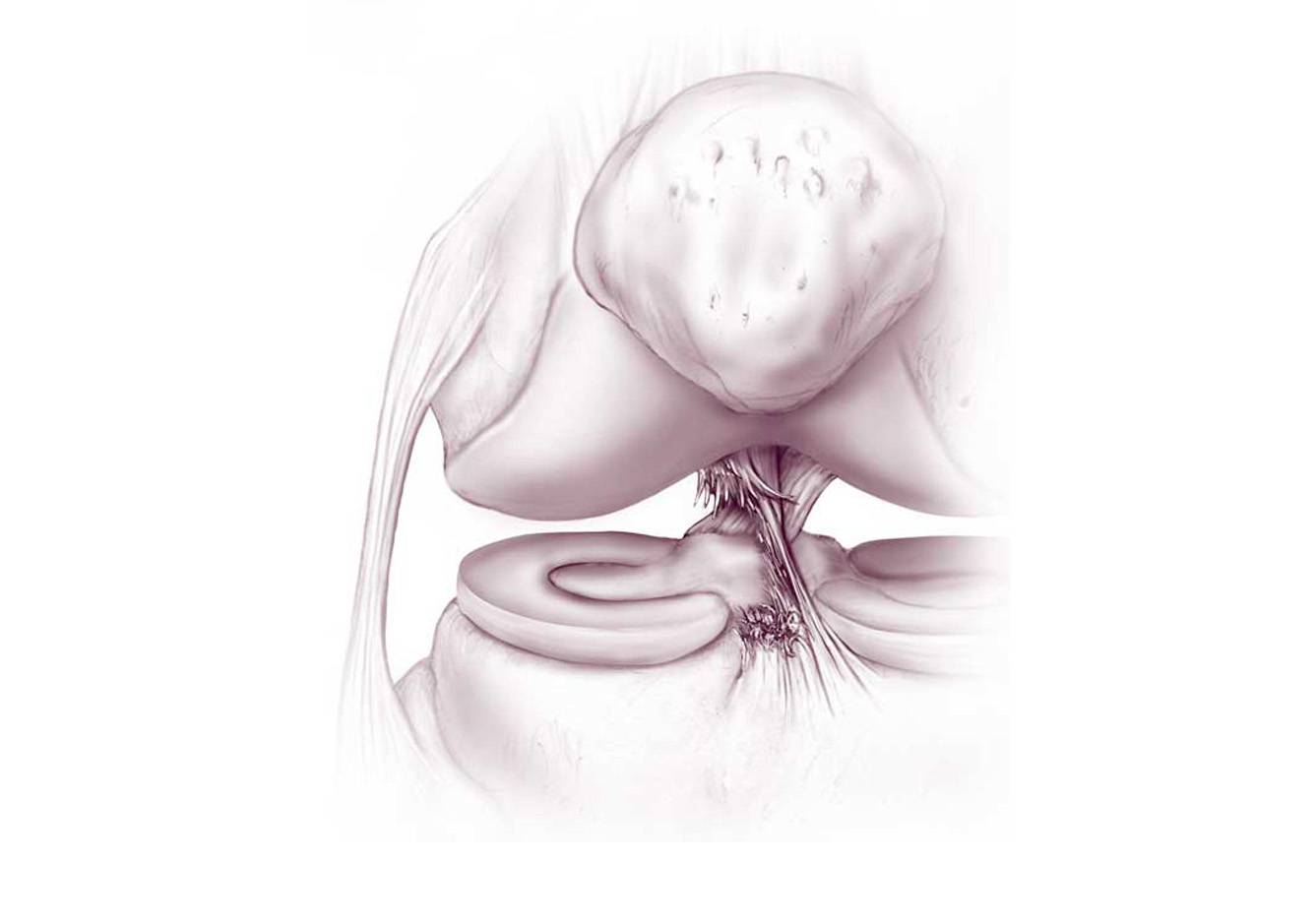
The cruciate ligaments form the "central pivot" of the knee, controlling the joint's alignment during movement. The collateral ligaments and menisci contribute to knee stabilization. These structures can also be damaged in the event of an ACL tear and must be repaired to restore full knee function.
Risk factors
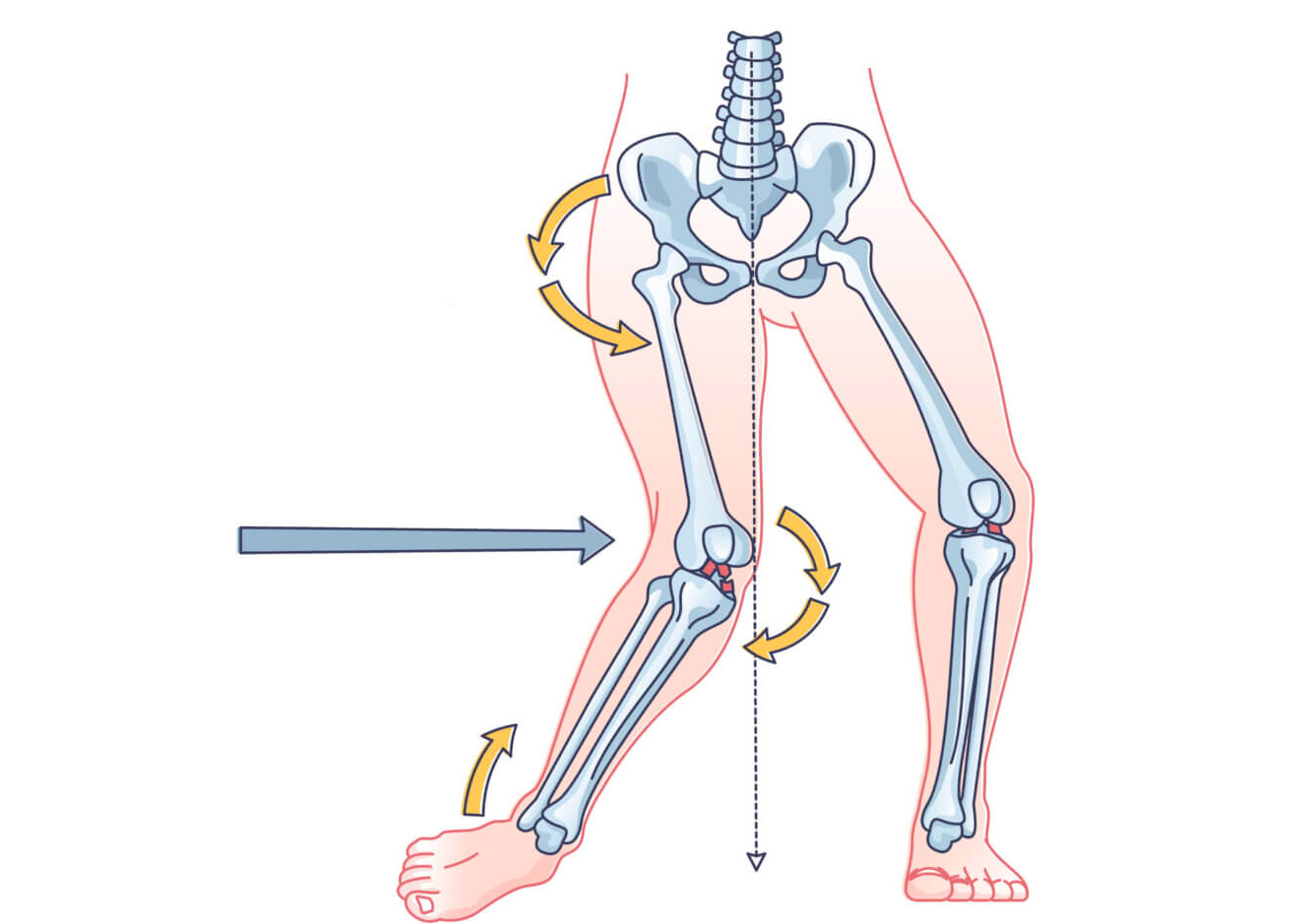
Risk factors are numerous and sometimes little known, such as Functional Hallux Limitus (www.fhl.science), which, through a sudden pronated tilt of the foot during a change of direction, causes the tibia to rotate internally and exposes the ACL to rupture. The activity performed also plays a significant role, such as ball sports where the ACL is more exposed. In skiing, the foot and ankle are immobilized, and the knee must bear additional stress, which is transferred to the ACL.
Poor physical condition, muscle fatigue, and balance are also risk factors that can lead to knee or ankle sprains. An unpredictable movement at the end of a game or day can cause a loss of knee control and result in an ACL injury.
Mechanism of injury
75 % Sprains occur during non-contact trauma. The injury mainly occurs during a sudden change of direction or landing off balance from a jump. The prognosis differs depending on whether the trauma is low-velocity or high-velocity (the latter being more frequent). The sprain occurs during a sudden deceleration combined with a change of direction that unbalances the knee and causes a dislocation of the tibia anterior to the femur, which tears the ACL. This sudden movement is due to a pronated foot roll that causes the knee to spiral into a valgus-internal tibial rotation, the "medial collapse" of the knee.
Other mechanisms exist, notably direct impacts, but these are much less frequent. Associated injuries, especially meniscal tears, are present in about half of cases and are treated simultaneously during surgery.
Diagnosis
Symptoms
The presence of significant fluid buildup, a popping sound at the time of the injury, or a feeling of instability with joint dislocation should raise suspicion of an ACL tear. Other symptoms include the need to stop activity due to pain, the inability to fully bend or straighten the knee, and a fear of instability while walking. In the hours following the injury, swelling of the knee frequently appears due to bleeding within the joint.
Clinical diagnosis
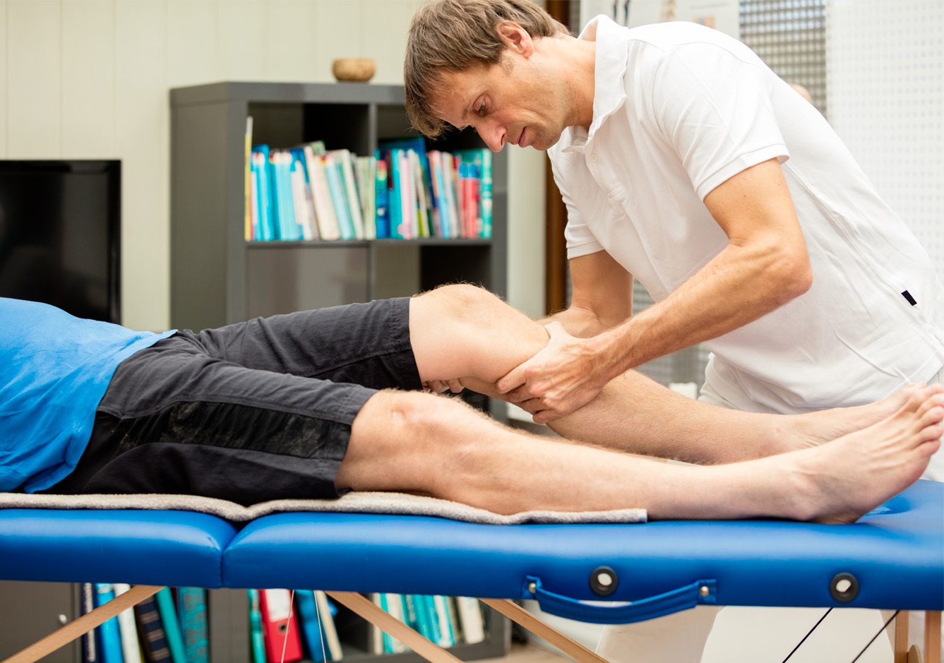
Clinical examination is sufficient in almost all cases to diagnose an ACL injury. This examination is sometimes difficult in emergency situations due to pain and muscle spasms around the knee. The presence of blood in the joint after a knee sprain indicates an anterior cruciate ligament tear in more than 70% of cases.
Reliable specific tests can confirm an ACL rupture. Two clinical maneuvers are specific: the anterior drawer test at 20° of flexion called the Lachman test, and the tibial pivot shift maneuver anterior to the femur called the pivot shift or Losee test.
Radiological diagnosis and MRI
Knee X-rays are usually normal, and an MRI is necessary to visualize the ACL tear. This examination also allows for the diagnosis of associated meniscal, ligament, or cartilage lesions. Meniscal tears are present in more than half of all anterior cruciate ligament tears.
Medical and sports context
Assessing individual functional abilities is essential for guiding treatment. This includes a standardized questionnaire, a mobility and stability assessment, and an evaluation of the patient's health status and sports activities. This assessment allows treatment to be tailored to each patient's specific needs and to define realistic recovery goals based on their expectations and abilities.
Preparation for surgery
Why is surgery necessary?
The ACL is a key element in the rotational stability of the knee. In the event of an injury, a feeling of insecurity may be experienced, such as a sensation of instability during certain movements or apprehension when walking (on uneven ground, going down stairs). Anterior cruciate ligament reconstruction is then necessary to eliminate this instability and restore confidence in the knee. The types of activities the patient wishes to resume after their ligament tear must also be taken into account.
In cases of associated meniscal tears, ACL reconstruction is necessary to ensure proper healing of these structures after repair. Meniscal surgery is a preservation procedure, not a resection, and must be performed relatively soon after the injury to achieve good results.
Knee inflammation and pain
Pain is usually relieved by medication, but if the knee is very swollen, the pain can be unbearable. In such cases, the knee must be drained under pressure to remove the fluid, and relief is immediate.
More than the ACL tear itself, it's often the associated injuries that are painful. A tear of the medial collateral ligament, in particular, can delay ACL reconstruction by several weeks because the pain can hinder rehabilitation. Therefore, it's important not to rush and to wait until the knee has fully recovered.
Walking with crutches is important because it helps relieve pain and allows you to walk without a limp. A knee brace may sometimes be prescribed for peripheral injuries, but its drawback is that it can disrupt the recovery of muscle control in the knee.
Active knee control
Before knee surgery, it is essential to have regained a normal, controlled gait, even if a slight extension deficit persists. The surgery involves reconstructing the ligament, not repairing it. Therefore, as it is an elective procedure, it is important not to rush into it unless serious meniscal or bone damage necessitates it.
The patient should not be left to their own devices, and physiotherapy is quickly initiated to maintain knee control and muscle strength. The program includes local treatment with lymphatic drainage massage, gait retraining, and active knee stabilization and muscle strengthening exercises.
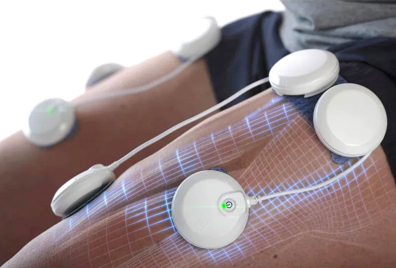
Particular attention is paid to the musculature, especially motor inhibition of the quadriceps muscle. This variable has recently become a major factor in surgical decisions. If you have motor inhibition, we can offer you specialized treatment, including electrostimulation.
The quality of your preparation and motivation are the best assets for facilitated rehabilitation and a return to your activities as soon as possible.
Operation
Ligament reconstruction
Unfortunately, repairing a cruciate ligament by suturing or bone reattachment is only possible in a very small percentage of cases. Most often, the tear is located deep within the ligament, and a simple suture is insufficient to guarantee knee stability. Ligament reconstruction is therefore necessary, and several techniques exist, using different grafts and fixation methods, specific to each surgeon.
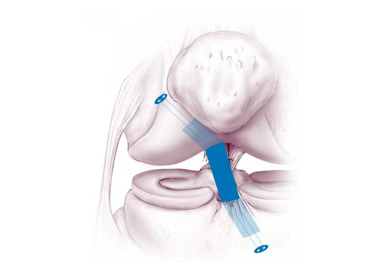
The principle of surgical ACL reconstruction involves rebuilding the anterior cruciate ligament with a tendon graft (autograft or allograft) that faithfully reproduces the tension and natural position of the anterior cruciate ligament. The operation is performed under arthroscopic guidance to ensure proper placement of the new ligament.
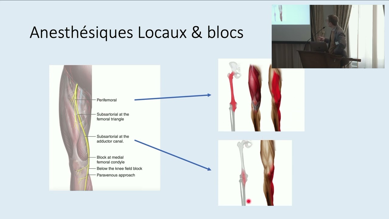
The procedure is performed under general or regional anesthesia (spinal anesthesia) with combined post-operative analgesia (nerve block and medication).
Treatment of meniscal tears
The menisci are essential for joint stability and load distribution during walking. Therefore, they are repaired in case of tearing, and the prognosis for healing is favorable when the ACL is reconstructed simultaneously.
Additional actions
Certain additional procedures may be performed, such as supplementary extra-articular surgery, realignment osteotomy, or correction of a dysfunction at a later stage (e.g., functional hallux limitus). Each case must be assessed individually to improve the prognosis.
Visit to the anesthesiologist
Before your procedure, you will meet with one of our anesthesiologists.
During this consultation, he will ask you questions about your health and perform a clinical examination.
You will also be able to discuss with him the type of anesthesia recommended and the measures put in place to ensure the best possible comfort after the operation.
Several solutions exist, all effective and tailored to your situation.
Rehabilitation and risks
Rehabilitation Principles
Rehabilitation following anterior cruciate ligament reconstruction takes into account the biological principles of tendon graft healing and the healing of associated injuries. Gait retraining begins on the day of surgery, initially with partial weight-bearing and then gradually with full weight-bearing. Each patient's progress is individual and should be respected as much as possible.
First days after the operation
Ice, lymphatic drainage, muscle activation exercises, and gait retraining are part of the program. Each person progresses at their own pace, according to their abilities and the treatment undertaken. Once walking on stairs is mastered, independence is regained, and pain is controlled, a return home is considered.
Return home
Upon your return, you will be able to continue the exercises demonstrated and practiced during your stay at the clinic, and you will walk outdoors for at least 20 minutes twice a day. You will have access to anti-inflammatory, pain-relieving, and anti-thrombotic medication, a prescription for physiotherapy (sessions should be taken two to three times a week), and an appointment with your surgeon for dressing changes, suture removal, and a clinical check-up, as well as a detailed rehabilitation program.
At home, the exercises should be performed at least twice a day in sessions lasting approximately one hour, with all exercises grouped together. The intensity of the exercises should be adjusted according to the swelling and pain in the knee. It is essential to walk, avoiding a limp, using crutches for this purpose.
The steps in your program
A detailed program of exercises and tests designed to ensure a smooth return to your activities will be sent to you weekly throughout your rehabilitation. You will receive exercises by email to help you practice daily, and any suggestions or comments you may have are welcome.
Infection
Infection is always a serious complication in orthopedic surgery, but fortunately very rare (less than 0.5 %). Specific measures are taken to minimize these risks: systematic pre- and immediate post-operative antibiotic therapy, operating rooms equipped with high-flow laminar airflow, a highly trained and experienced orthopedic surgical team, optimal skin preparation, and careful monitoring of your health. If you develop a fever or local redness after your procedure, once you have returned home, you must contact your surgeon immediately.
Venous thrombosis and pulmonary embolism
Venous thrombosis is characterized by the blockage of a vein by a clot (thrombus), which prevents blood from returning to the heart. If the clot migrates, it can in turn obstruct the pulmonary vessels, resulting in a pulmonary embolism. To minimize these risks, a tourniquet will not be used during your procedure. Prevention will be achieved through subcutaneous injections of low molecular weight heparin, and gait rehabilitation will begin as soon as possible. Antithromboembolic prophylaxis will be prescribed for five to ten days following your procedure. If you experience calf pain or tenderness during the postoperative period at home, you must contact your surgeon immediately.
Nerve damage
It is possible that you may experience decreased sensation in the upper (proximal) part of your tibia after surgery, due to stretching of a superficial nerve branch. The incision is made near a sensory branch, which can be stretched during the procedure. Sensation usually returns after several weeks. More rarely, a neuroma can cause persistent pain.
Functional limitation
The knee may retain some stiffness after surgery and not regain its full range of motion. This can be due to muscle weakness, the formation of scar tissue, or adhesions within the knee. Treatment should be preventative and is now well-established.
Medicol Specifics
Active rehabilitation
The Medicol rehabilitation program aims to inform you and empower you to participate with the healthcare team in the success of your procedure. This program is based on close support during the pre-hospitalization phase, during your stay at the clinic, and upon your return home.
The main goal of this program is for you to feel confident throughout your rehabilitation and for the steps set for your progress to seem safely achievable. Find your cruising speed and progress at your own pace and without pain with the help of your therapist.
Your journey
After your consultation with the surgeon, during which the operation is scheduled, a follow-up consultation with a resident physician is planned to discuss the information received and ensure everything is clearly understood and organized. You will then have the opportunity to ask any remaining questions. During this consultation, a questionnaire will be added to your file along with specific data from the clinical examination. The purpose of collecting this information is to assess your expectations and correlate them with your abilities to ensure optimal and personalized care.
Back to sport
Returning to sports is a gradual process, both at the end and during rehabilitation. Beforehand, it's essential to regain control of the knee through increasingly demanding activities and rebuild good muscle strength. Throughout your physiotherapy treatment, several progress tests will be conducted to monitor your development and allow you to progress at your own pace.
Activities in which you feel comfortable are prioritized. The main goal is to regain a smooth, balanced, and limp-free gait. Keep using your crutches long enough to be able to walk without a limp by the time you decide to stop using them. Letting go of the crutches isn't a challenge; your friends will help you find your footing again and learn to walk correctly.
Returning to sports is a gradual process: it begins with resuming cycling, cross-country skiing, and Nordic walking after six weeks. Two and a half months after the procedure, a specific training program can already be offered depending on the sport practiced. Before resuming pivot-contact sports (starts, sudden stops, contact, and changes of direction) such as badminton, squash, tennis, football, basketball, ice hockey, skiing, etc., we recommend a gradual return-to-sport phase using mime exercises: returning to the sidelines to repeat sequences of your sport, without contact, at progressively increasing speeds.
Frequently Asked Questions
What kind of scar will I have?
What sports will I be able to do after my procedure?
How long will I need to wear a splint?
How long will I need to use crutches?
How long does the healing process take to integrate the new ligament?
When will I be able to take a shower?
When will I be able to drive?
Summary

Need medical advice?
The doctors at the Ouchy Orthopedic Center are at your service.
1006 Lausanne
+41 21 510 33 48
Learn more in videos
Medicol Videos
17:46
16:25
26:04
12:00
15:00
17:07
16:28
1:19:31
Contact one of our specialists on the subject
Ouchy Orthopedic Center
Specialized medical consultations in orthopedics and traumatology in the heart of Lausanne

 Français
  English (UK)