
 English

- Welcome
- Shoulder osteoarthritis
Shoulder osteoarthritis
Shoulder
Introduction
Anatomy
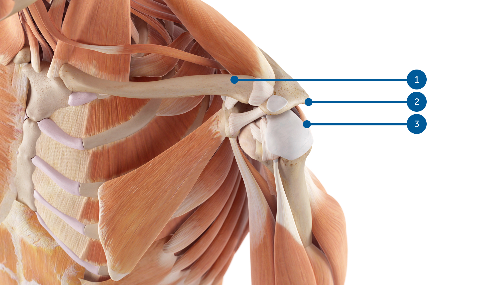
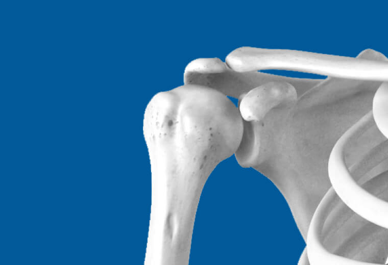
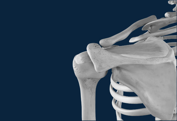
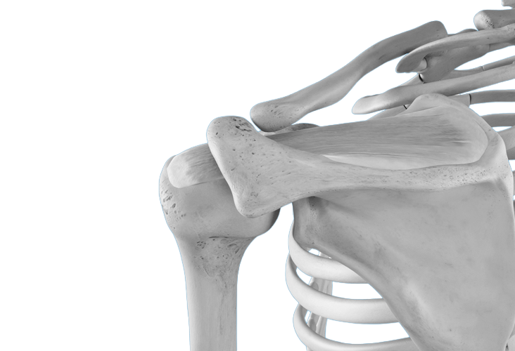
(1) Clavicle, (2) Acromion, (3) Supraspinatus (4) Capsule, (5) Subscapularis (Subscapularis)
The shoulder is remarkable for its range of motion in all directions thanks to the joints that compose it:
- Sternoclavicular joint
- Acromioclavicular joint
- Glenohumeral joint
- Scapulothoracic joint (sliding of the shoulder blade on the thorax).
To animate the shoulder and at the same time stabilize it by pressing the articular surfaces against each other, there is a set of very effective small muscles grouped under the term "rotator cuff" because of their anatomical arrangement above the head of the humerus.
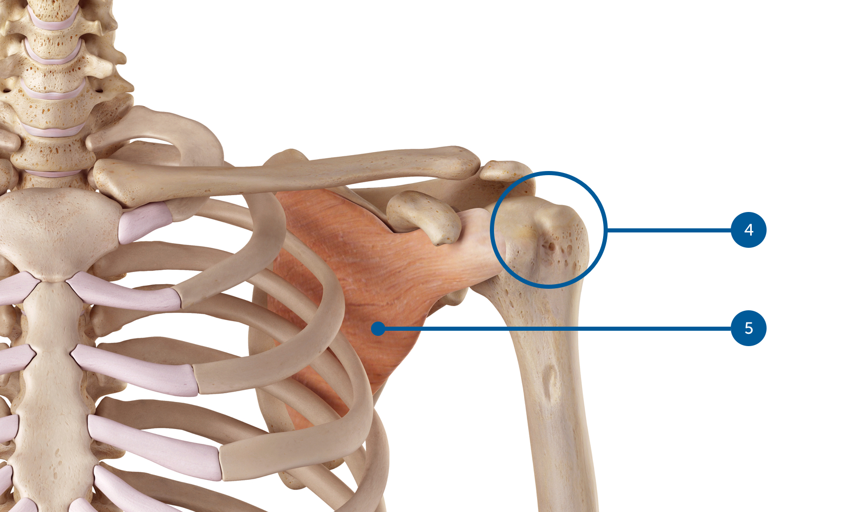
These rotator muscles of the shoulder are:
- the supraspinatus (Supraspinatus)
- the infraspinatus
- the subscapularis
- the small circle (Teres Minor).
The supraspinatus also plays a key role in raising the arm and allows the head of the humerus to slide under the bony roof of the shoulder called the acromion, which is part of the scapula.
Signs & symptoms
The main symptom of shoulder osteoarthritis is pain, associated with joint stiffness. The pain is present when the shoulder is moved and frequently wakes the patient at night. In cases of associated rotator cuff tears, these symptoms may be accompanied by muscle weakness. As osteoarthritis is a progressive disease, an increase in pain and a gradual decline in shoulder function are frequently observed over time.
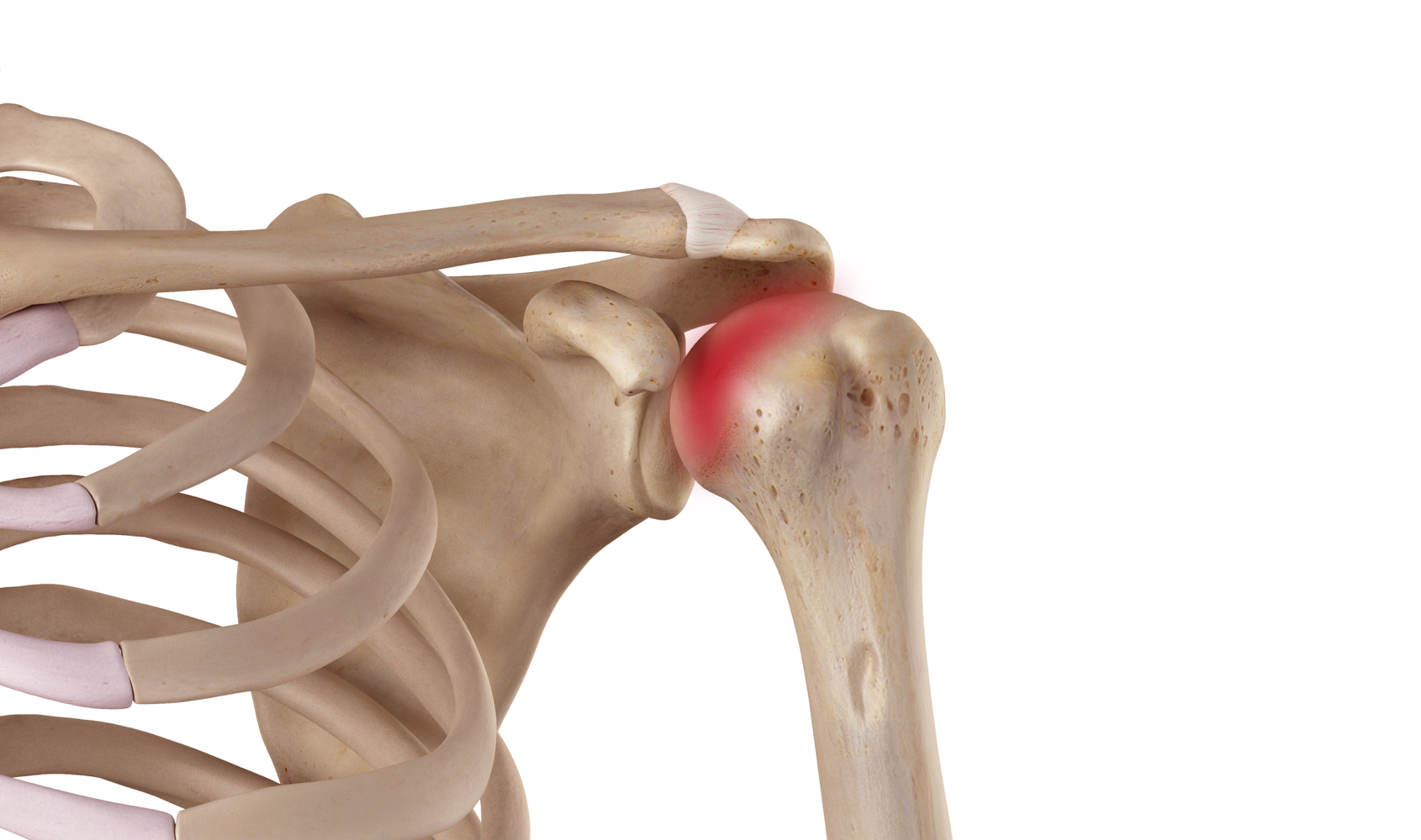
What is this?
The goal of prosthetic shoulder treatment is to relieve pain and improve shoulder function.
Shoulder surgery may be necessary due to various conditions that damage the cartilage and/or tendons of the rotator cuff. This damage can result from several pathologies:
- Rheumatic arthritis (glenohumeral osteoarthritis or rheumatoid arthritis)
- Avascular necrosis of the humeral head
- Chronic and irreparable rupture (massive muscle atrophy) of the rotator cuff
- Fractures or sequelae of fractures of the humeral head
The principle of the surgery is to replace the damaged joint surfaces with prosthetic implants.
A total shoulder replacement is used when the injury involves the humeral head and the glenoid cavity. The replacement will be performed using a humeral component and a glenoid cup.
- A shoulder hemiprosthesis is used when the injury only affects the humeral head. The replacement will be performed using only the humeral component.
- A reverse total shoulder replacement is used when there are associated rotator cuff tears. The design of this prosthesis compensates for tendon insufficiency.
Risk factors
- Repetitive microtrauma (activities that put repetitive strain on the shoulder)
- Joint injuries (fractures, chronic rotator cuff tears)
- Microcrystalline and rheumatic diseases (rheumatoid arthritis, chondrocalcinosis, etc.)
- Heredity
- Chronic shoulder instability
Screening & Diagnosis
Clinical examination is essential: shoulder mobilization and specific tests to assess rotator cuff functionality.
Standard radiological examination of the shoulder can identify osteoarthritis. Additional imaging with MRI and CT scans, combined with arthrography, allows for the evaluation of the rotator cuff and bone stock in preparation for prosthesis implantation.

Treatments
Self-treatment
Resting the joint and taking anti-inflammatory medication can relieve pain. In early-stage osteoarthritis, a cortisone injection and a well-conducted physiotherapy program can reduce symptoms.
Osteoarthritis is a progressive disease, and an increase in pain and a gradual decline in shoulder function are frequently observed over time. If the pain persists, it is best to consult a doctor.
Medical treatments
The type of treatment is tailored to the severity of the osteoarthritis, its functional impact, and the pain.
Physiotherapy treatment aims to reduce inflammation and pain. It is based primarily on loosening the capsular ligaments, relaxation, and then muscle strengthening.
Since the effect of conservative treatment is limited in advanced osteoarthritis, surgical management represents the most appropriate therapeutic solution.
Our favorite methods
The procedure is performed under general anesthesia combined with a nerve block for pain relief. The catheter will be removed after 2-3 days, once the pain has subsided.
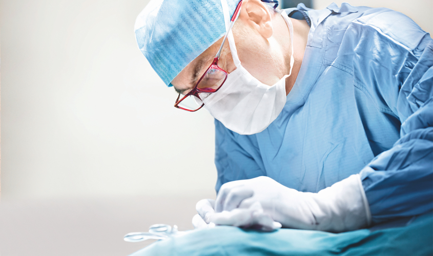
The procedure requires a skin incision of approximately 10 cm on the front of the shoulder. The operation is performed in a sterile environment with laminar flow, and under prophylactic antibiotic protection.
The choice of prosthesis depends on the integrity of the rotator cuff, bone stock, and functional requirements. The average surgery lasts approximately 1 hour and 30 minutes. The arm is then immobilized in an orthopedic sling for 4 to 6 weeks.
Post-operative rehabilitation follows a protocol. The hospital stay is approximately one week. Assistance from the community health center (CMS) for meals, personal hygiene, and housekeeping can be arranged upon request after discharge.
Surgery
Preparation for surgery
We are available to inform your primary care physician about the proposed treatment so that we can provide you with the best possible care, prevent risk factors, and ensure optimal postoperative follow-up. Once the surgical treatment is scheduled, your primary care physician may be asked to complete sections of a form that will be sent to the anesthesiologist. This request is standard practice for major procedures.
An appointment with the anesthesiologist may be deemed useful or essential depending on the type of procedure and any existing risk factors. This appointment takes place before or upon your admission to the clinic, or even on the day of the operation itself, for example. The purpose of this consultation is to provide you with all the necessary information regarding the type of anesthesia, which will be decided upon in consultation with you. The discussion will cover not only the anesthesia itself but also methods for managing pain during the postoperative period.
Depending on the surgical schedule, you will be admitted the day before your procedure or on the same day, 3 hours before the operation, on an empty stomach.
Recovery
A rehabilitation program begins the day after surgery. Physiotherapists will mobilize the operated arm and teach exercises to be performed independently. Physiotherapy must be continued on an outpatient basis.
The hand and elbow can be moved at the waist, protected by an orthopedic vest for six weeks. The orthopedic vest can be removed daily during the day for washing and dressing. Since the shoulder cannot be moved above the torso, driving is not permitted during these six weeks.
Possible risks
Complications following shoulder replacement surgery are rare. Here are the most common ones, for which we implement active prevention measures.
Shoulder stiffness. A rehabilitation program can usually treat it.
The infection. It is treated with antibiotics and may sometimes require removal of the prosthesis and its replacement at a later stage.
Dislocation. It is treated by surgical revision when it recurs.
Neurological and vascular damage is extremely rare, but is always a possible complication regardless of the type of surgery performed.
Wear and tear (loosening) of the prosthesis generally occurs late, after more than 10 years. It is sometimes necessary to replace the implants.
Frequently Asked Questions
What type of anesthesia is offered for this type of surgery?
How long should I wear a scarf or vest?
Is surgical treatment often synonymous with success?
When will I be able to start driving again?
Summary

Need medical advice?
The doctors at the Ouchy Orthopedic Center are at your service.
1006 Lausanne
+41 21 510 33 48
Learn more in videos
Medicol Videos
20:36
15:29
Contact one of our specialists on the subject
Ouchy Orthopedic Center
Specialized medical consultations in orthopedics and traumatology in the heart of Lausanne

 Français
  English (UK)